
Kidney Microanatomy (Lesson) Human Bio Media
A nephron is the unit of structure and function in the kidney. Each nephron is a coiled tube held together by a tough fibrous connective tissue. In humans, a healthy adult has 1 to 1.5 million nephrons in each kidney, functioning together to filter blood from all its impurities. They also regulate blood pressure, control electrolytes, and.

How To Draw Nephron step by step for beginners YouTube
#nephron #howtodraw #adimushowThis is an easy and simple drawing of the nephron. This will teach you how to draw the nephron diagram easily. This is a step-b.
Draw a diagram of nephron and explain its structure.
Each nephron has basically two parts.. 'Nephron Diagram || How to draw and label the parts of a Nephron' is demonstrated in this video tutorial step by step.
Nephron Definition, Parts, Structure, & Functions, with Diagram
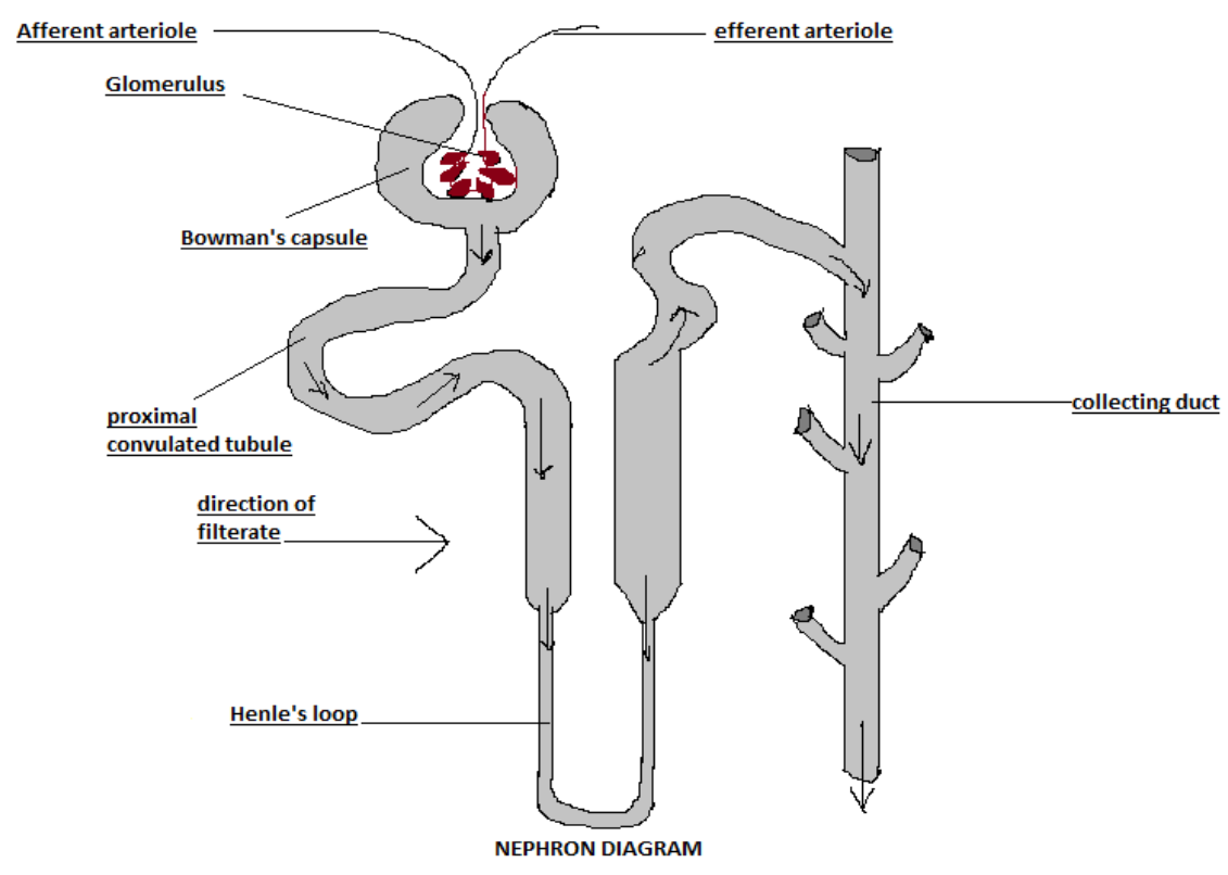
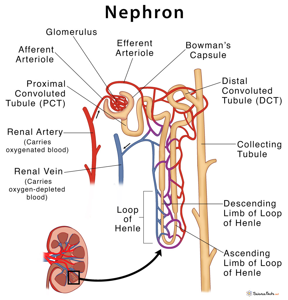
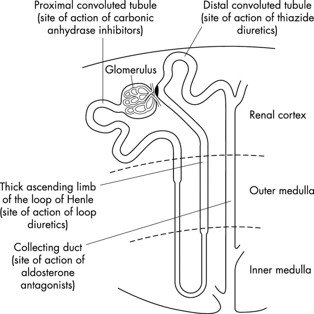
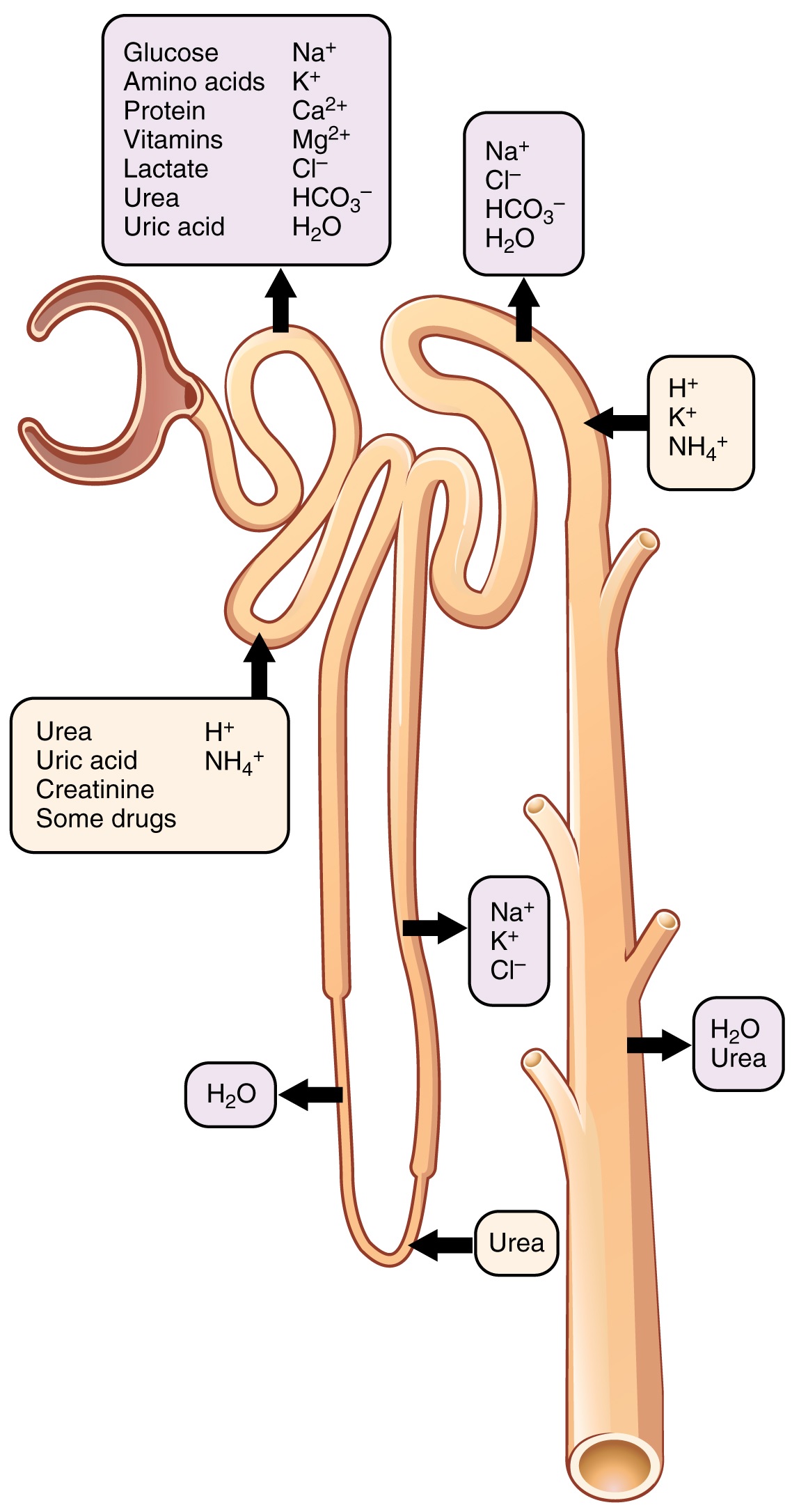
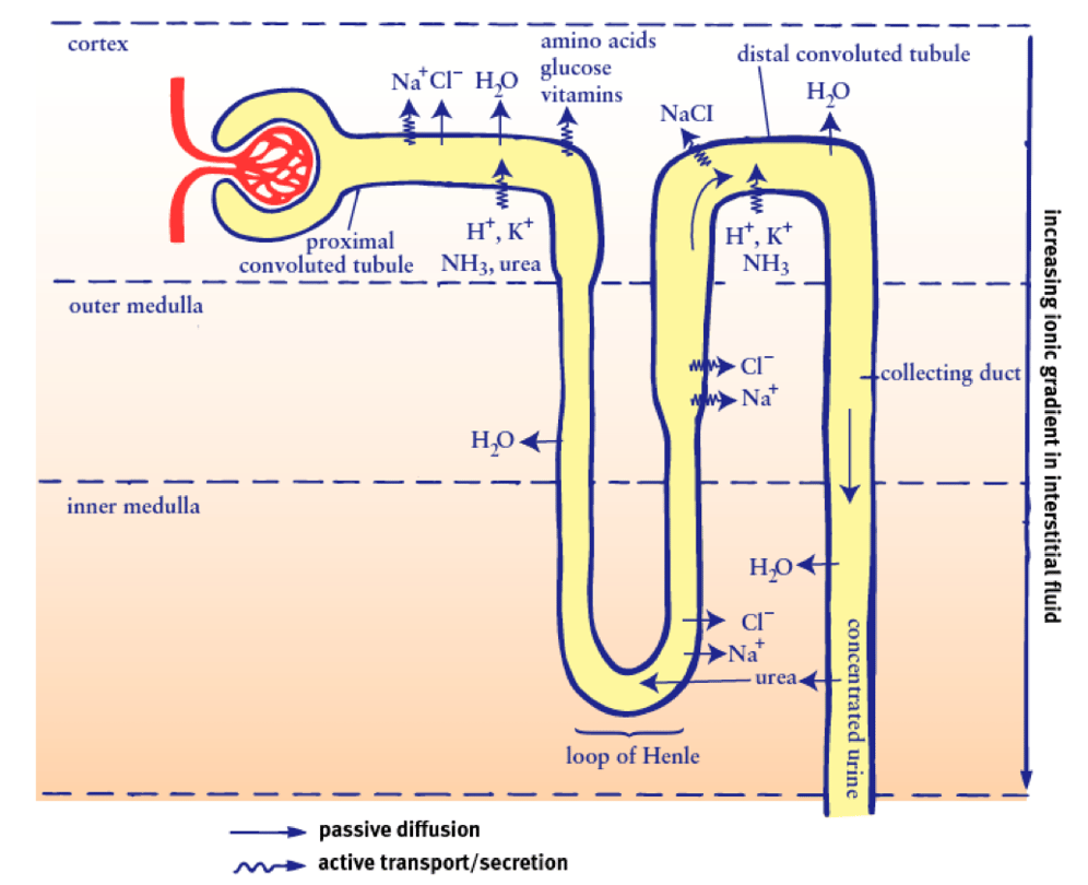
Nephron (overview) Ultrastructurally, the nephron is the functional representative of the kidney. Each nephron contains arenal corpuscle, which is the initial component that filters the blood, and a renal tubule that processes and carries the filtered fluid to the system of calyces. The renal corpuscle has two components: the glomerular (Bowman.

Simple Structure Of Nephron Diagram bmpmongoose
A nephron is the basic unit of structure in the kidney. A nephron is used separate to water, ions and small molecules from the blood, filter out wastes and toxins, and return needed molecules to the blood. The nephron functions through ultrafiltration. Ultrafiltration occurs when blood pressure forces water and other small molecules through.

Structure Of A Nephron Formation Of The Urine Stock Illustration Download Image Now Nephron
Diagram of the Nephron, Glomerulus and Different Parts of the Tubule. The tubules are lined with a thin layer of epithelial cells. The thinner segments of the tubules like the descending limb and first part of the ascending limb of the loop of Henle has cells with minimal organelles. The other segments have thicker cells with a brush border and.

Schematic drawing of a nephron. Illustration printed with permission... Download Scientific
The nephron of the kidney in mammals is a tube that is approximately 30-55 mm in length. The nephron has an inflated and closed tube. The end of this tube is folded to a cuplike shape structure.
Nephron The Functioning Unit of The Kidney Interactive Biology, with Leslie Samuel
This video explains How to draw Nephron in easy steps and compact way. This video helps you to draw science diagrams with great ease and clarity.Nephron is t.
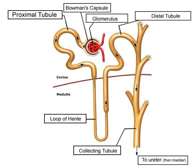
Diagram of nephron Healthiack
Below is a well-labelled and easy diagram of the nephron for your better understanding. Broadly, a nephron can be divided into two parts - renal corpuscle and renal tubule. The renal corpuscle is the filtering component and renal tubules carry the filtered liquid away. A human nephron is a long fine tubule that is about 30-55 mm long.

Diagram of the nephron segments and its juxtaglomerular apparatus Download Scientific Diagram
The purpose of this study is to see whether a large drawing of a nephron helped medical students in self-directed learning groups learn renal physiology, histology, and pharmacology before discussing clinical cases. The end points were the grades on the renal examination and a student survey. The classes in the fall of 2014 and 2015 used the.

How To Draw Structure Of Nephron Diagram Step By Step For Beginners YouTube
The nephron is the minute or microscopic structural and functional unit of the kidney.It is composed of a renal corpuscle and a renal tubule.The renal corpuscle consists of a tuft of capillaries called a glomerulus and a cup-shaped structure called Bowman's capsule.The renal tubule extends from the capsule. The capsule and tubule are connected and are composed of epithelial cells with a lumen.

How to draw easy diagram of nephron step by step for beginners YouTube
This video tutorial demonstrates how to draw Nephron step by step. You can learn how to draw and label the parts of Nephron diagram, just watching this vide.
Structure and Functions of Nephron
1/4. Synonyms: Cortex renalis. The kidneys are paired retroperitoneal organs of the urinary system. Their function is to filter blood and produce urine. Each kidney consists of a cortex, medulla and calyces. The nephron is the main functional unit of the kidney, in charge of removing metabolic waste and excess water from the blood.
CC Breaking down nephron functioning into six easy steps!
A nephron is the basic structural and functional unit of the kidneys that regulates water and soluble substances in the blood by filtering the blood, reabsorbing what is needed, and excreting the rest as urine. Its function is vital for homeostasis of blood volume, blood pressure, and plasma osmolarity. It is regulated by the neuroendocrine.

Nephron What is the Structure and its Functions of Nephron?
How to draw nephron in easy steps : Life Processes | CBSE Class 10 Science Syllabus | Biology | NCERT 10th Class | Best Animated video Lectures | grade boost.

Label The Diagram Of The Kidney And Nephron Below Free Diagram For Student
The purpose of this study is to see whether a large drawing of a nephron helped medical students in self-directed learning groups learn renal physiology, histology, and pharmacology before discussing clinical cases. The end points were the grades on the renal examination and a student survey. The classes in the fall of 2014 and 2015 used the drawing, but not those of 2012 and 2013. The Charles.
